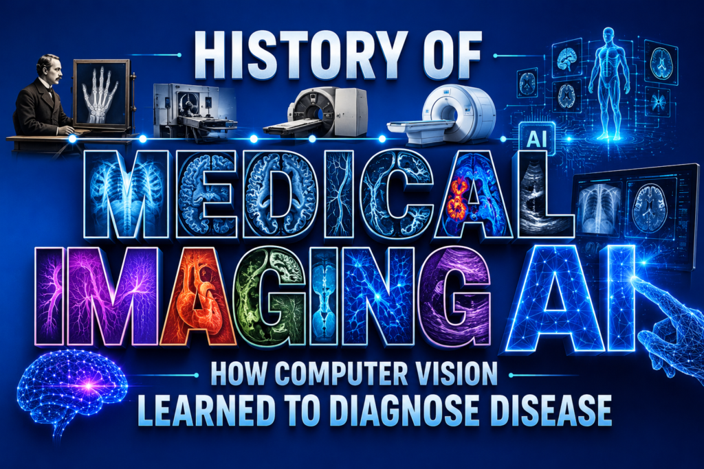
The journey of medical imaging ai is an incredible story of technological triumph. Healthcare has always relied on the sharp eyes of doctors to find hidden ailments within the human body. Over the last several decades, scientists have worked tirelessly to teach machines how to interpret these complex visuals. By understanding the Evolution of AI in medical imaging, we can appreciate the immense leaps made in healthcare technology. From rudimentary algorithms to sophisticated neural networks, the ability of computers to see and analyze has fundamentally reshaped patient care. The integration of this technology promises faster results and life saving accuracy. The transformation did not happen overnight. It took generations of brilliant computer scientists, radiologists, and engineers working together to bridge the gap between silicon chips and organic biology. Today, we stand on the precipice of a new frontier in medicine where algorithms are indispensable diagnostic partners.
The Dawn of Digital Discovery (1960 – 1970)
Before algorithms could identify tumors, researchers had to figure out how to translate visual data into numbers. The history of image processing reveals that early computers struggled with even the simplest shapes. Scientists in the mid twentieth century experimented with rule based systems to help interpret crude radiological scans. These pioneering efforts laid the necessary groundwork for future innovations. While modern medical imaging ai feels like magic, its roots are firmly planted in these early attempts to quantify shadows and contrast. The machines of this era lacked the computing power we take for granted today, yet the vision of automated diagnosis was born.
During this decade, the very concept of digitizing a medical photograph was revolutionary. Engineers had to develop the hardware to capture images and the software to convert light intensities into binary code. Early research focused on basic edge detection and pixel intensity thresholding. These rudimentary techniques were slow and prone to errors, but they proved that a machine could theoretically look at an anatomical structure. The scientific community recognized the massive potential, even if the practical applications were still decades away.
Rise of Expert Systems (1970 – 1990)
As hospitals began adopting new scanning technologies, the sheer volume of data exploded. The invention of Computed Tomography (CT) scans provided unprecedented three dimensional views of human anatomy. Suddenly, doctors were not just looking at flat images; they were navigating through slices of the human body. However, interpreting these slices required immense expertise. During this period, the history of pattern recognition took center stage as computer scientists developed expert systems. These programs attempted to mimic the decision making processes of human specialists by using complex logical frameworks.
The History of automated MRI scanning diagnostics also traces back to this era, when researchers first tried to extract measurable features from magnetic resonance signals. While early programs were rigid and often failed outside controlled laboratory settings, they proved that machines could assist in identifying abnormalities. This era highlighted the need for more flexible learning algorithms that could adapt to the immense variability in human biology, a fundamental requirement for true medical imaging ai. A major roadblock was the limitation of hand crafted rules. A programmer had to explicitly tell the computer what a tumor looked like, specifying its shape, size, and density. But human biology is wildly unpredictable. Tumors rarely follow strict geometric rules. Consequently, these early expert systems suffered from high false positive rates, frequently flagging normal tissue as pathological. It became clear that the software needed to learn from experience rather than relying solely on programmed instructions.
The Era of Machine Learning (1990 – 2010)
The transition from rigid rules to systems that learn from data marked a massive turning point. The FDA approved AI medical imaging software history officially began in the late nineteen nineties with tools designed to rescreen Pap smears. This was a monumental milestone, validating the commercial and clinical viability of automated screening tools. Around the same time, the medical community established DICOM format standardization, ensuring that images from different machines could be shared and analyzed uniformly. This crucial step paved the way for larger datasets, which are the lifeblood of machine learning.
Researchers started exploring Medical image analysis using deep neural networks, although hardware limitations still restricted their potential. Convolutional neural networks in digital pathology began to show promise for analyzing tissue samples at microscopic levels, identifying cellular structures that were invisible to the naked eye. The foundation for robust medical imaging ai was solidifying, as algorithms became better at filtering noise and highlighting regions of interest for human doctors. The shift toward statistical learning allowed algorithms to weigh probabilities rather than making absolute binary decisions. By analyzing thousands of historical cases, the software could identify subtle correlations between pixel arrangements and disease outcomes. This decade laid the intellectual groundwork for the explosion of artificial intelligence that was about to follow.
The Deep Learning Boom (2010 – 2020)
Everything changed when deep learning transformed computer vision in the early twenty tens. Powerful graphics processing units allowed scientists to train massive models on thousands of images simultaneously. The introduction of the U-Net segmentation model revolutionized how computers outline organs and anomalies. Instead of just flagging a suspicious area, the software could now draw precise boundaries around complex structures. By leveraging transfer learning in computer vision, developers could take models trained on everyday objects and adapt them for clinical use. Transfer learning from ImageNet became a standard practice, significantly reducing the time and data required to build effective medical tools.
During this decade, Deep learning image segmentation for tumors became incredibly precise, rivaling human experts in certain specialized tasks, propelling medical imaging ai into the mainstream. As algorithms became smarter, Computer vision applications in radiology expanded rapidly across the globe. The history of image segmentation reached a pinnacle as systems learned to separate overlapping structures with pinpoint accuracy, distinguishing between blood vessels, bone, and soft tissue seamlessly. Hospitals began deploying Clinical decision support systems (CDSS) to aid radiologists during their gruelling shifts. These tools acted as a second pair of eyes, ensuring that subtle fractures or tiny nodules were not overlooked amid the chaos of a busy emergency room. The integration of medical imaging ai moved from theoretical research into daily clinical practice, saving lives through earlier and more accurate detections. Furthermore, cloud computing enabled smaller clinics to access cutting edge analytical tools without investing in expensive local servers, democratizing access to high quality diagnostics.
Modern Era and Complex Workflows (2020 – 2030)
Today, the capabilities of medical imaging ai are truly breathtaking. Engineers are tackling incredibly complex problems like 3D volumetric pixel (Voxel) processing to analyze whole organs in real time. Instead of looking at flat slices sequentially, modern software evaluates the entire organ as a cohesive three dimensional entity. One of the primary goals of modern systems is False negative rate reduction, ensuring that dangerous conditions are never missed. Computer vision for early cancer detection is now a reality, with algorithms spotting malignant patterns years before they become visible to the naked eye. Radiomics feature extraction allows software to pull thousands of invisible data points from a single scan, predicting patient outcomes with astonishing accuracy. This means doctors can tailor treatments based on the specific microscopic characteristics of a patient’s tumor.
However, the field still faces significant hurdles that require collaborative solutions. The Challenges of medical imaging AI datasets remain a constant topic of debate among researchers and ethicists. Training these massive models requires millions of high quality examples representing diverse demographics. Relying heavily on Semantic annotations by radiologists is expensive and time consuming, as every single image must be meticulously labeled by an expert before the computer can learn from it. Furthermore, ensuring Generalization across scanner hardware is critical, because an algorithm that works perfectly on a machine in New York might fail completely on a different brand of scanner in Tokyo. Variations in image contrast, resolution, and noise profiles can easily confuse poorly trained models. Despite these obstacles, developers continue to refine Deep learning models for X-ray analysis and other modalities, pushing the boundaries of what is medically possible. Regulatory bodies are also adapting, creating new frameworks to evaluate software that continuously learns and updates itself over time.
FAQs About medical imaging ai
What exactly is medical imaging ai?
It refers to the use of artificial intelligence algorithms to analyze scans like X rays and MRIs. The goal is to assist doctors in diagnosing diseases more quickly and accurately than traditional manual methods. By processing massive amounts of visual data instantly, these tools provide valuable insights that improve overall clinical workflows.
How does it improve patient care?
By highlighting subtle anomalies, medical imaging ai helps catch conditions like cancer or fractures at their earliest stages. This leads to faster treatment plans and generally better outcomes for the patients involved. It also reduces the burden on overworked healthcare professionals, minimizing the risk of diagnostic fatigue.
Will machines replace human radiologists?
No, medical imaging ai is designed to augment human expertise rather than replace it. It handles repetitive tasks and acts as a safeguard, allowing doctors to focus on complex cases and direct patient care. The combination of human intuition and machine precision yields the best possible results.
Is patient data safe when using these AI tools?
Yes, strict privacy laws and robust encryption methods protect patient data. When images are used to train new models, they are thoroughly anonymized to ensure that personal identities are never revealed or compromised. Hospitals utilize highly secure servers to process and store this sensitive information safely.
Conclusion
The fascinating history of medical imaging ai showcases human ingenuity at its finest. What began as simple experiments in contrast adjustment has evolved into sophisticated networks capable of identifying life threatening diseases in milliseconds. As we look to the future, the continued refinement of computer vision technology will undoubtedly unlock even greater potential in healthcare. The journey from rudimentary pixel counting to profound diagnostic insight has revolutionized how we approach medical science. By combining the empathy and contextual knowledge of human doctors with the relentless precision of artificial intelligence, the medical community is poised to deliver a level of care that was once considered impossible. The evolution of medical imaging ai is far from over, and its next chapter promises to be even more extraordinary for patients worldwide.